What is Fibrocartilaginous Embolism (FCE)?
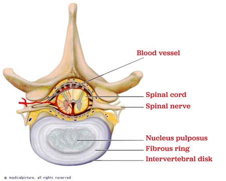
The intervertebral discs are located between the bones of the spine (the vertebrae), and these intervertebral discs contain a substance called fibrocartilage. A small piece of this fibrocartilage can exit the intervertebral disc and make its way into the blood vessels that supply the spinal cord. This piece of fibrocartilage can then block the normal blood flow to an area of the spinal cord, which means that oxygen and other nutrients cannot reach that part of the spinal cord. This ultimately leads to damage to the cells of the spinal cord. This is called a Fibrocartilaginous Embolism (v), and you can think of it as a “stroke” to the spinal cord.
What Clinical Signs Occur with FCE?
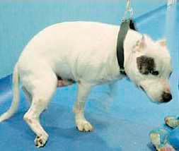
The most common sign of an FCE is a sudden onset of weakness in one limb, both hind limbs, or all four limbs. An uncoordinated, spastic, or wobbly gait may also be seen, and some animals may stand with their paw(s) knuckled over. In severe cases, paralysis (inability to move) can occur. An FCE is not usually a painful process, however, some dogs and cats may be painful for the first 24 hours. The onset of clinical signs is usually very sudden, and commonly occurs during normal exercise or activity. Most dogs and cats with an FCE will have a short period of progressive neurologic dysfunction, but in the majority of cases the neurologic abnormalities do not worsen after the first 24 hours.
Which Animals are Prone to Developing FCE?
Any animal can develop an FCE, but some dogs and cats are more prone to developing this disease than others. A fibrocartilaginous embolism most commonly occur in young to middle-aged large breed and giant breed dogs. Miniature Schnauzers and Shetland Sheepdogs are the most commonly affected small breed dogs. Middle-aged to older cats are more commonly affected, and Domestic Shorthair cats are the most common cat breed affected by an FCE.
How is FCE Diagnosed?
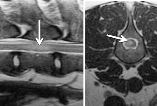
MRI of the spinal cord is gold standard way to diagnose an ANNPE, and doing this test can help us rule-out other causes of your pet’s spinal cord dysfunction (like a compressive intervertebral disc, a fibrocartilaginous embolism, a tumor, or an infection). Usually, the MRI will show an area of increased signal (brightness) within the spinal cord directly over the intervertebral disc space.
For some dogs and cats, a spinal tap will be recommended to rule-out the presence of inflammation or infection of the spinal cord. The spinal fluid is sent to a laboratory for analysis. Most animals with an ANNPE have an increased protein level in their spinal fluid.
How is FCE Treated?
There is no specific treatment for an ANNPE, and no medications (including high dose steroids) have been proven to help speed the recovery from an ANNPE. Surgery is not helpful since the spinal cord is not compressed by this jelly-like nucleus pulposus.
The only treatment which has been proven to help speed the recovery from an ANNPE is physical therapy. Physical therapy is strongly recommended for most dogs and cats recovering from an ANNPE.
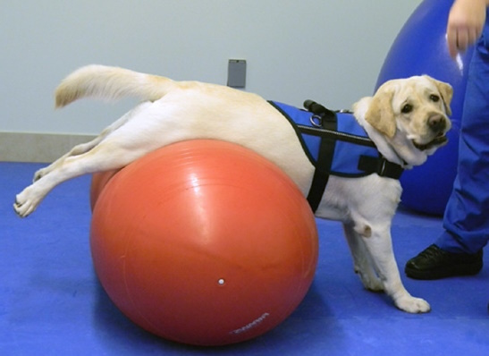
There is no specific treatment for an FCE, and no medications (including high dose steroids) have been proven to help speed the recovery from an FCE. The only treatment which has been proven to help speed the recovery from an FCE is physical therapy. Physical therapy is strongly recommended for most dogs and cats recovering from an FCE.
Nursing care is very important for animals recovering from an FCE, especially those who cannot move their limbs or urinate on their own.
If your pet cannot move well on their own, you will need to rotate the side they are laying on every 4 to 6 hours to prevent bedsores. It’s also very important to make sure your pet has soft, clean, and dry bedding to lie on, which will also prevent bedsores. During your pet’s recovery, it is important to keep them away from slippery surfaces or stairs where they could fall and injure themselves.
If your pet is dragging their toes or knuckling their paws over, you will need to inspect their feet twice daily to check for ulcers or sores. It is a good idea to keep them away from rough surfaces like pavement or asphalt. You can place a bootie or sock on your pet’s feet to help prevent the development of sores.
There are booties specifically made for dogs and cats, and these can be purchased here, at most pet stores, or online. If you notice sores or wounds on your pet’s feet, please contact us or your veterinarian. If your pet cannot walk on their own, a sling or harness is very useful to help support their weight. There are slings and harnesses specifically made for dogs and cats, and these can be purchased here, at most pet stores, or online.
Some pets may require help with urination during their recovery. This may require you to express their bladder or to administer medications to help make urination easier.
What is the Prognosis for Fibrocartilaginous Embolism?
The prognosis for recovery from an FCE varies, and it is usually dependent on the severity of the neurologic dysfunction. Dogs or cats that lose the ability to feel their toes (loss of deep pain sensation), tend to have a poorer prognosis for recovery. In one study of 50 dogs with an FCE, 84% of dogs were able to return to walking and had a good quality of life. In one study of 19 cats with an FCE, 79% of cats recovered the ability to walk. The prognostic value of an MRI is vital as certain characteristics can help predict both positive and negative outcomes. Most dogs and cats begin to show signs of neurologic improvement within the first 2 weeks. While recurrence of an FCE is possible, this is extremely rare.
References
- Theobald A, Volk AH, Dennis R, et al. Clinical outcome in 19 cats with clinical and magnetic resonance imaging diagnosis of ischaemic myelopathy (2000-2011). JFMS 2013; 15(2), 132-141.
- De Risio L, Adams V, Dennis R, et al. Association of clinical and magnetic resonance imaging findings with outcome in dogs suspected to have ischemic myelopathy: 50 cases (2000- 2006). JAVMA 2008; 233, 129-135.
- Mikszewki JS. Fibrocartilaginous embolic myelopathy in five cats. JAAHA 2006; 42, 226-233.
- Dewey CW. Myelopathies: Disorders of the Spinal Cord. A Practical Guide to Canine and Feline Neurology, 2008:369-372.
- Platt S, Garosi L. Ischaemic Myelopathy. Small Animal Neurological